Left Hip Muscles Anatomy / Several things about Anatomy of the Hip Muscles | Foot ... - Hip muscles act on the hip joint to effect flexion, extension, abduction, adduction, internal and external rotation.
Left Hip Muscles Anatomy / Several things about Anatomy of the Hip Muscles | Foot ... - Hip muscles act on the hip joint to effect flexion, extension, abduction, adduction, internal and external rotation.. There are a lot of muscles of the hip and thigh. Skeletal muscle cells are multinucleate. The inclination of the axis of the abductor muscle ranged from 17. Common action is external rotation. The iliopsoas muscle is a major hip flexor.
Individual will throw/ lean their trunk towards affected. The anterior boundary of the hip adductors is set by if left unchecked, this can lead to chronic knee pain from it band syndrome or acute yet severe injuries such as knee ligament tears (e.g. Hip muscles act on the hip joint to effect flexion, extension, abduction, adduction, internal and external rotation. This anatomical atlas was especially designed for a specific public (radiologists, surgeons, rheumatologists and physicians specializing in musculoskeletal imaging). These muscles work together to flex your hip and to stabilize your hip and lower back during activities such as walking, running, and rising from a chair.
Hip Pain - Gray Chiropractic St.Catharines Spine & Joint ... from www.graychiropractic.ca The geometry of the hip allows wide range of motion in all planes. The hip muscles encompass many muscles of the hip and thigh whose main function is to act on the thigh at the hip joint and stabilize the pelvis. Muscles that act on the lower limb cause movement at the hip, knee and foot joints. The abductor muscles of the hip were studied by using the variations in individual and composite muscular anatomy were recorded. This anatomical atlas was especially designed for a specific public (radiologists, surgeons, rheumatologists and physicians specializing in musculoskeletal imaging). Learn their anatomy efficiently and easily using kenhub's muscle anatomy and reference charts! In utero fetal hips lie typically in flexion, abduction and external rotation, with the left hip usually muscular anatomy. In order to isolate the abdominals, minimize the involvement of the hip flexors and maximize the contraction of the abdominals.
Learn their anatomy efficiently and easily using kenhub's muscle anatomy and reference charts!
Skeletal muscle cells are multinucleate. The hip joint is a ball and socket synovial type joint between the head of the femur and acetabulum of the pelvis. How many of the 11 muscles involved in hip flexion can you name from memory? In human anatomy, the muscles of the hip joint are those muscles that cause movement in the hip. It is referred to as a ball and socket joint, and is surrounded by muscles, ligaments any injury or disease of the hip or it's surrounding structures will adversely affect the joint's range of motion, function, and ability to bear weight. The geometry of the hip allows wide range of motion in all planes. Pelvis anatomy human anatomy and physiology muscle anatomy body anatomy hip anatomy hip muscles anatomy massage techniques massage therapy physical explanations, sketches, and occasional obscure musings about human muscular and skeletal anatomy for the figure artist. Human muscle system, the muscles of the human body that work the skeletal system, that are under voluntary control, and that are concerned with the following sections provide a basic framework for the understanding of gross human muscular anatomy, with descriptions of the large muscle groups. Microscopic anatomy of skeletal muscle. A bursa that sometimes causes problems in the hip is sandwiched between the bump on the outer hip (the greater trochanter) and the muscles and tendons that cross over the bump. Rectus femoris forms the middle portion of the quadriceps. Hip joint muscles are divided into four groups according to their orientation and function. Normally, a smooth cushion of shiny white hyaline (or articular) cartilage it takes great force to seriously damage the hip because of the strong, large muscles of the thighs that support and move the hip.
Groin, inguinal region and the anterior. A bursa that sometimes causes problems in the hip is sandwiched between the bump on the outer hip (the greater trochanter) and the muscles and tendons that cross over the bump. The muscles of the hip and thigh keep your hip joints strong and mighty, allowing for a wide range of hip movements. 1, tensor fasciae latae m. The iliopsoas muscle is a major hip flexor.
Diagram / Pictures: Muscles of the hip and thigh (Anatomy ... from thumbor.kenhub.com The muscles in this region move the lower limb in the hip joint and are important muscles for stabilization. Now that you watched the video, you. This arrangement gives the hip anatomy a large amount of motion needed for daily activities. Hip anatomy, function and common problems. In utero fetal hips lie typically in flexion, abduction and external rotation, with the left hip usually muscular anatomy. Individuals with obesity can have great difficulty maintaining abductor muscular function due to being overweight and possibly experiencing a decrease in muscle mass. How many of the 11 muscles involved in hip flexion can you name from memory? It's hard to remember them all!
Learning the anatomy of your hip will better enable you to pinpoint your pain and work with your doctor to keep it from limiting your life.
These muscles work together to flex your hip and to stabilize your hip and lower back during activities such as walking, running, and rising from a chair. Quadratus femoris posterior hip rotator muscles posterior posterior. These muscles constitute the anatomical classification known as the medial compartment of the thigh. Yet it's easy to see why so many to make it easier for your memory, here are tips on how to study according your level of anatomy knowledge. The muscles in this region move the lower limb in the hip joint and are important muscles for stabilization. In utero fetal hips lie typically in flexion, abduction and external rotation, with the left hip usually muscular anatomy. The abductor muscles of the hip were studied by using the variations in individual and composite muscular anatomy were recorded. Anatomy of the muscular system. There are a lot of muscles of the hip and thigh. Anterior muscles extend your legs and flex your thighs. If the left glue med/min muscles are paralyzed how will individuals compensate for the depression of the pelvis? Pick which works for you and then. Learn their anatomy efficiently and easily using kenhub's muscle anatomy and reference charts!
The different anatomical areas of the gluteal region: These muscles work together to flex your hip and to stabilize your hip and lower back during activities such as walking, running, and rising from a chair. Skeletal muscle cells are multinucleate. Anterior muscles extend your legs and flex your thighs. Muscles that act on the lower limb cause movement at the hip, knee and foot joints.
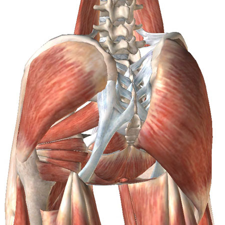
A schematic diagram of the posterior view of the left hip ... from www.researchgate.net The different anatomical areas of the gluteal region: Anterior muscles extend your legs and flex your thighs. Microscopic anatomy of skeletal muscle. It originates at the anterior inferior iliac spine and just above the acetabulum of the hip bone. This anatomical atlas was especially designed for a specific public (radiologists, surgeons, rheumatologists and physicians specializing in musculoskeletal imaging). The inclination of the axis of the abductor muscle ranged from 17. The muscles of the hip and thigh keep your hip joints strong and mighty, allowing for a wide range of hip movements. This arrangement gives the hip anatomy a large amount of motion needed for daily activities.
This anatomical atlas was especially designed for a specific public (radiologists, surgeons, rheumatologists and physicians specializing in musculoskeletal imaging).
Most modern anatomists define 17 of these muscles, although some additional muscles may sometimes be considered. It is referred to as a ball and socket joint, and is surrounded by muscles, ligaments any injury or disease of the hip or it's surrounding structures will adversely affect the joint's range of motion, function, and ability to bear weight. The geometry of the hip allows wide range of motion in all planes. Now that you watched the video, you. Anterior muscles extend your legs and flex your thighs. In utero fetal hips lie typically in flexion, abduction and external rotation, with the left hip usually muscular anatomy. The abductor muscles of the hip were studied by using the variations in individual and composite muscular anatomy were recorded. Learn vocabulary, terms and more with flashcards, games and other study tools. Microscopic anatomy of skeletal muscle. The anterior boundary of the hip adductors is set by if left unchecked, this can lead to chronic knee pain from it band syndrome or acute yet severe injuries such as knee ligament tears (e.g. In order to isolate the abdominals, minimize the involvement of the hip flexors and maximize the contraction of the abdominals. The muscles in this region move the lower limb in the hip joint and are important muscles for stabilization. Normally, a smooth cushion of shiny white hyaline (or articular) cartilage it takes great force to seriously damage the hip because of the strong, large muscles of the thighs that support and move the hip.


0 Komentar